Stationary digital breast tomosynthesis increases diagnostic accuracy
26 Mar 2019
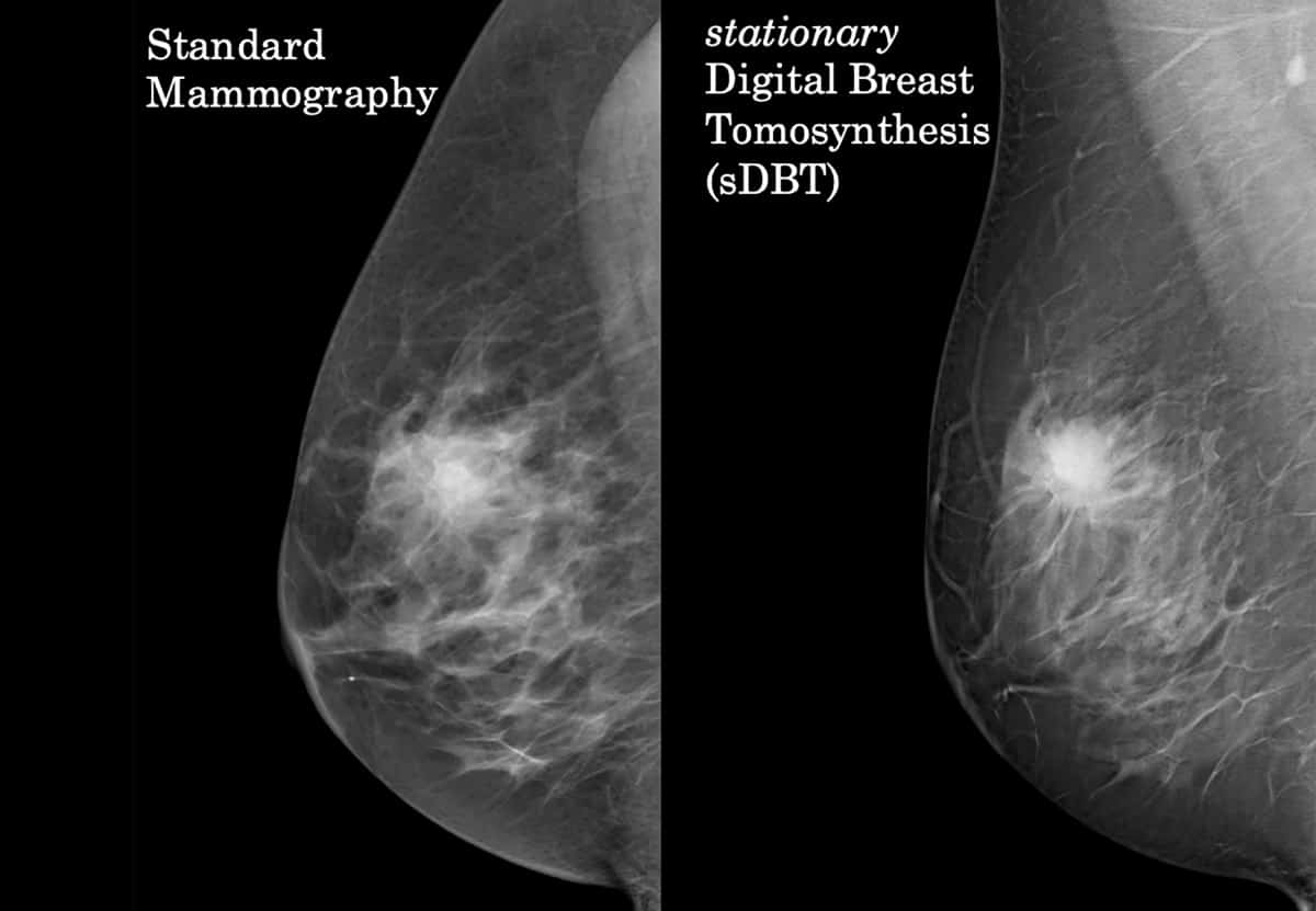
The addition of digital breast tomosynthesis (DBT) to a 2D mammography exam can significantly improve breast cancer detection by making lesions more conspicuous on a pseudo-3D image. DBT has limitations, however, including image degradation due to focal spot blurring, noise, scatter and motion artifacts, as well as the inability to visualize breast microcalcifications as well as conventional mammographic images.
To overcome these shortcomings, researchers are developing stationary DBT (sDBT) devices. Existing commercial DBT systems work by moving a single X-ray tube — either by continuous motion or a step-and-shoot technique — to collect a series of projection views at multiple angles. In sDBT, the single rotating X-ray tube is replaced by a fixed array of carbon nanotube-enabled (CNT) X-ray sources. This allows for a rapid, motion-free collection of multiple projection views over a wide-angle span. The stationary DBT system. (Courtesy: Yueh Lee)
 The stationary DBT system. (Courtesy: Yueh Lee)
A team at the University of North Carolina are developing an sDBT device based on a modified commercial DBT system (Selenia Dimensions) in which a fixed array of 15 CNT-enabled X-ray sources replaces the standard X-ray source. Each source consists of a CNT cathode, a gate, and an individual tungsten anode. The CNT cathodes release electrons at room temperature in response to an applied voltage, allowing for precise X-ray production on demand. The researchers have now published results from their initial clinical evaluation of this first-generation sDBT system (Acad. Radiol.10.1016/j.acra.2018.12.026).
Lead author Yueh Lee and colleagues conducted a paired-image study, evaluating mammography and sDBT images of 43 women with suspicious findings identified on a prior mammogram. The subjects included 28 patients with dense breasts. The average compressed breast thickness was 4.7 cm for sDBT, and 4.6 cm for mammography. Twelve of the patients had breast cancer, six with infiltrating ductal carcinomas, five with intraductal carcinomas and one with invasive lobular carcinoma.
The scan time to acquire 15 projection images using sDBT was less than 5 s for both craniocaudal and mediolateral oblique views, with a radiation dose equivalent to that of a conventional tomosynthesis scan. The researchers reconstructed the image slices using a thin depth increment of 0.5 mm to ensure that small features were displayed sharply.
Four radiologists with 10 to 25 years of experience reading mammograms and up to five years interpreting DBT assessed the images. For each exam, they were asked to rate the likelihood of malignancy in increments of 10%, as well as the confidence of their overall impression. They also used BIRADS A-D classification to record the density of breast tissue.
After reading both sets of images, the radiologists rated their preference for the set of images presented first — either mammography or sDBT — when assessing diagnostically important image features. They repeated this exercise at least four weeks later, first interpreting the images from the modality that they had reviewed last.
On average, the radiologists were more likely to identify a malignancy correctly when interpreting the sDBT images. This higher diagnostic accuracy held true for each breast density category and breast
thickness range. Only the most experienced radiologist, with 25 years of mammography experience, performed better when reading mammograms. The readers preferred sDBT when interpreting soft-tissue features, including mass shape and margins, architectural distortion and asymmetry. As expected, they preferred mammography images for identification and characterization of microcalcifications.
“The team’s goal is to continue to improve resolution and reduce imaging time through further tube development,” Lee tells Physics World. “We are also exploring improvements in synthetic 2D mammograms based on our technology. Our hope is that the sDBT can completely eliminate the need for the 2D mammography shot, and reduce breast compression time, in addition to all the benefits of conventional breast tomosynthesis.”
“In addition to advancing woman’s imaging, our collaborative team is working hard to develop novel, low-dose clinical applications of the carbon nanotube X-ray source, including chest, cardiac, dental, orthopedic and brain imaging,” Lee adds.
The researchers also plan to investigate the effects of slice thickness on the visibility of microcalcification clusters with sDBT. They also are considering measuring the relationship between experience with both modalities and level of training with respect to sDBT interpretation performance.
Cynthia E Keen is a freelance journalist specializing in medicine and healthcare-related innovations
FROM PHYSICSWORLD.COM 30/3/2019
Δεν υπάρχουν σχόλια:
Δημοσίευση σχολίου