Near infrared fluorescence imaging provides early diagnosis of cracked teeth
10 Jul 2020
Cracked teeth can be identified in their early stages using near infrared fluorescence (NIRF) imaging, researchers in the US have demonstrated. The approach – which can distinguish between different types of crack and reveal their depth – is more reliable than existing modalities and may help better diagnose the source of otherwise inexplicable toothache.
Cracked teeth are a common condition and, thanks to their potential to allow bacteria across the enamel–dentin junction, also the third highest cause of tooth loss. Despite this, the condition is often overlooked in its early stages. “Cracked teeth can be difficult to diagnose clinically as patients’ symptoms often aren’t reproducible and cracks can be barely visible to the naked eye,” explains oral surgeon James Allison of Newcastle University, who was not involved in the present study.
At present, there is no dependable clinical method for detecting the presence of cracks in tooth enamel. Visual and surgical-microscope-aided inspection is an unreliable approach, and dye-staining is time consuming and cannot reveal cracks beneath the surface of teeth. Common imaging modalities like X-ray and cone-beam CT, meanwhile, do not offer a high enough resolution. MicroCT scanning, with its higher resolution, can reveal larger cracks – but is only viable on extracted teeth, rendering it useless in a clinical setting.
Conventional near-infrared imaging – in which light is passed through dental structures and scattered to produce image contrast – has, like X-ray imaging, been shown capable of detecting some cracks in teeth. But it cannot distinguish between crack types or provide further information on the extent of the damage.
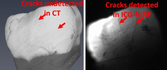
In their study, engineer Jian Xu of the Louisiana State University and colleagues have instead turned to NIRF – in which image contrast is generated by the differential accumulation within teeth of a fluorescent dye (here indocyanine green) which is excited by infrared light. To demonstrate the potential of the technique, the researchers compared the images of 16 extracted cracked teeth produced by NIRF with both those from near-infrared transillumination and X-ray imaging.
They found that the fluorescence approach was consistently able to reveal cracks in enamel that were not visible in the X-ray images – and was able to highlight more cracks than conventional near infrared imaging. They also report that an angled exposure gave better image contrast, as it created shadows under each crack. From these, one can determine crack depth and whether the crack is in enamel alone or has also reached the dentin.
Furthermore, the team noted that cracks could be revealed by immersing teeth in the fluorescence agent for only one minute – although longer periods produced clearer images. In practice, the dye could be applied to patient teeth via a mouthwash. In fact, indocyanine green has the benefit of being entirely safe to swallow – although has been known to cause allergic reactions.
“We use indocyanine-green-assisted near-infrared imaging to address the major drawback of the current state-of-the-art dental imaging: failure to detect some critical dental diseases, for example, early stage cracks and caries,” Xu tells Physics World. Alongside this, he adds, the new technique does not rely on the use of bulky imaging sensors and avoids the ionizing radiation-based health risks associated with X-ray techniques.
“The idea of checking all teeth for cracks would be unlikely to be cost effective as a health intervention,” notes dental radiologist Keith Horner of University Dental Hospital Manchester. Enamel cracks are not normally treated, he explains, but the approach could be useful for diagnosing patients with toothache where decay or restoration is not an obvious cause of the pain.
The research is described in the Annals of the New York Academy of Sciences.
from physicsworld.com 15-07-2020

Δεν υπάρχουν σχόλια:
Δημοσίευση σχολίου