Hydrogel helps grow new tissue in areas of brain damage
10 Mar 2023 Tami Freeman
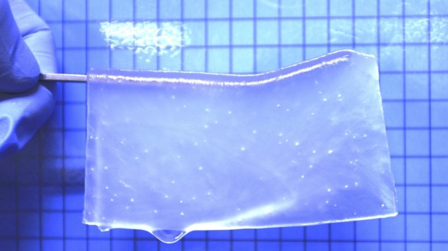
Healing the brain Researchers at Hokkaido University have created an optimized hydrogel material for brain tissue reconstruction. (Courtesy: Satoshi Tanikawa et al Scientific Reports)
Brain haemorrhage and brain cancer are major causes of death and disability worldwide. The brain is particularly vulnerable to ischemic damage, in which loss of blood supply leads to the loss of brain tissue volume and the formation of a cavity. And unlike other parts of the body where spontaneous wound healing processes occurs, loss of neuronal tissue is irreversible.
The development of an efficient technique for brain tissue regeneration is urgently needed to help treat these life-threatening conditions. To date, however, there is no established therapeutic strategy. One potential solution could lie in neural tissue engineering – the use of biomaterials to create a scaffold that fills the volume loss, enabling cells to migrate into the damaged area and reconstruct tissue.
Researchers at Hokkaido University have now created synthetic hydrogels that provide an effective scaffold for neuronal tissue growth in areas of brain damage. Describing the study in Scientific Reports, the team showed that the hydrogels, used in combination with neural stem cells (NSCs), could grow new brain tissue, providing a potential approach for brain tissue regeneration.
Hydrogel optimization
To create the ideal substrate for brain tissue reconstruction, Satoshi Tanikawa and colleagues altered the electric charge of the hydrogel to determine the conditions under which NSCs can efficiently attach and grow. They found that a neutral hydrogel with a 1:1 mixture of anionic and cationic monomers (C1A1) generated the most suitable scaffold for attachment, growth and differentiation of NSCs.
The researchers used cryogelation to create pores in the C1A1 hydrogels and enable 3D culture of cells in the porous structure. They also adjusted the ratios of crosslinker molecules to achieve a similar stiffness to that of brain tissue, an important parameter as the stiffness of the cell culture substrate can impact the direction of stem cell differentiation.
After optimizing the hydrogel formulation, the researchers examined its potential for in vivo brain tissue reconstruction. They created a model of traumatic brain injury in mice by removing a 1 mm-diameter, 1 mm-deep region of brain tissue, and then implanted the porous hydrogel into the cylindrical cavity.
On day 56 after hydrogel implantation, the boundary between the brain tissue and the hydrogel was obscured, while cavities clearly remained in control brains. Fluorescence microscopy revealed that immune cells and astrocytes from surrounding brain tissue had infiltrated into the pores of the implanted hydrogel. Fluorescence image Visualization of neurons and glial cells in the hydrogel. (Courtesy: Satoshi Tanikawa et al Scientific Reports)
 Fluorescence image Visualization of neurons and glial cells in the hydrogel. (Courtesy: Satoshi Tanikawa et al Scientific Reports)
For effective brain tissue reconstruction, creation of a vascular network in the new tissue is critical. To induce vascularization, the researchers immersed the C1A1 hydrogel in vascular endothelial growth factor (VEGF) before implantation into the mice brains. In vivo imaging using two-photon microscopy revealed that this step led to the formation of blood vessels inside the hydrogel two to three weeks after implantation
After three weeks, when blood vessels had formed and host glioneuronal cells had migrated, the team injected fluorescently labelled NSCs into the implanted hydrogel in the mouse brain. Forty days later, fluorescence images demonstrated the presence of viable stem cells in the hydrogels, with a high cell survival rate. Some of the NSCs had differentiated into new glial or neuronal cells, and some of the new neuronal cells had migrated into surrounding host brain tissue.
The researchers point out that direct injection of NSCs alone or NSCs with control Matrigel did not support cell survival in the mouse brain. They also highlight the importance of the two-step process – implanting the hydrogel and NSCs at the same time proved unsuccessful. Finally, they note that they did not see any behavioural abnormalities or deaths in any of the mice implanted with the hydrogels.READ MORE

“We demonstrated that C1A1 electrically charged porous hydrogels can serve as scaffolds for brain parenchymal defects, and stepwise transplantation of NSCs into the hydrogel following gel implantation may induce the reconstruction of brain tissue along with the implanted hydrogels,” the team writes. “Therefore, this two-step method for neural regeneration may become a new approach for therapeutic brain tissue reconstruction after brain damage in the future.”
“We are currently trying to prove functional recovery with our method, using a mouse model of brain damage,” senior author Shinya Tanaka tells Physics World. “This evaluation is very critical for future possible clinical applications.”

Tami Freeman is an online editor for Physics World
FROM PHYSICSWORLD.COM 16/3/2023

Δεν υπάρχουν σχόλια:
Δημοσίευση σχολίου