PET imaging tracks ingested microplastics in mice
27 Jul 2021
Microplastics, tiny pieces of plastic debris less than five millimetres in length, are designed for commercial use or created through the breakdown of consumer products and industrial waste. They litter our oceans, they have been detected in everything from aquatic life to drinking water, and they take lifetimes or longer to decompose. In 2019, the World Health Organization called for more research on the effects of microplastics to the environment and human health.
Humans ingest and inhale microplastics, and they also can absorb them through the skin. Less is known about the risks and health effects of these exposures. In some animal studies, exposures to large amounts of microplastics have been associated with inflammation, metabolic disruptions, increased cancer risk and other adverse health effects.
To begin to learn more about how microplastics interact with the body, researchers in South Korea turned to functional imaging.
Tracking radiolabelled microplastics in mice
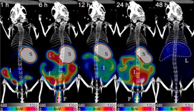
Jin Su Kim, Choong Mo Kang and their teams at the Korea Institute of Radiological & Medical Sciences and the University of Science and Technology observed and imaged mice that had ingested microplastics that were labelled, or paired, with a harmless radioactive compound.
The mice ate small plastic compounds joined to copper-64, a positron-emitting radioisotope, and DOTA, a molecule that helps bind copper-64 to plastic. For two days the researchers traced the paths taken by the tagged microplastics after they were ingested, by detecting pairs of gamma rays created during the decay of copper-64.
Positron emission tomography (PET) scans and accompanying gamma camera scans confirmed that the microplastics had spread throughout each animal’s body. Over the 48-hour observation period, the plastics passed through and were taken up by the stomach, intestines, liver, spleen, heart, lung, kidney, bladder and other organs. The researchers also observed the accumulation of radiolabelled copper-64 in tissues.
Ex vivo thin layer radio-chromatography confirmed that the small PET signals were indeed coming from radiolabelled microplastic and not another form of copper-64, and organs were identified using simultaneously acquired computed tomography (CT) scans.
What now?
The researchers’ results, which were published in the Journal of Nuclear Medicine, “may be used as the basis for future studies on the toxicity of microplastics,” says Kim, one of the senior authors on the study.
“There are many studies on the absorption of microplastics using fluorescent plastics in the body,” explains Kim. “However, we are the first to identify the in vivo absorption pathway of microplastics over time using radioisotope and PET techniques.”READ MORE

The big advantage of the researchers’ PET imaging technique is that, unlike fluorescent plastics-based studies, it doesn’t require animal sacrifices at each observation time point.
The researchers’ next steps are to evaluate the biological effects of long-term exposure to microplastics in organs and continue to identify the in vivo pathways of small pieces of plastic derived from the breakdown of larger plastic debris, another type of plastic that they would like to track with PET. Major challenges for future work include labelling radioisotopes to these larger microplastics, which requires the addition of molecules not ordinarily present in plastic, and then detecting useful signals.
Catherine Steffel is a science writer based in Madison, Wisconsin. Catherine was previously a PhD student contributor to Physics World.
from physicsworld.com 15/9/2021

Δεν υπάρχουν σχόλια:
Δημοσίευση σχολίου