PETRUS hybrid device acquires multimodal images in vivo
19 Oct 2018

A research team in France has evaluated the PETRUS hybrid imaging instrument both in vitroand in vivo in small animals. The researchers demonstrated the capability of the device, which simultaneously performs PET/CT and ultrafast ultrasound, to acquire multimodal images in vivo without significant degradation of image quality (Phys. Med. Biol. 63 19NT01).
The researchers — based at Inserm, the Université Paris Descartes and ESPCI Paris — explored the effect of using a PETRUS system on image quality, finding deviations of below 10% between images acquired with and without ultrasound probes.
Image quality
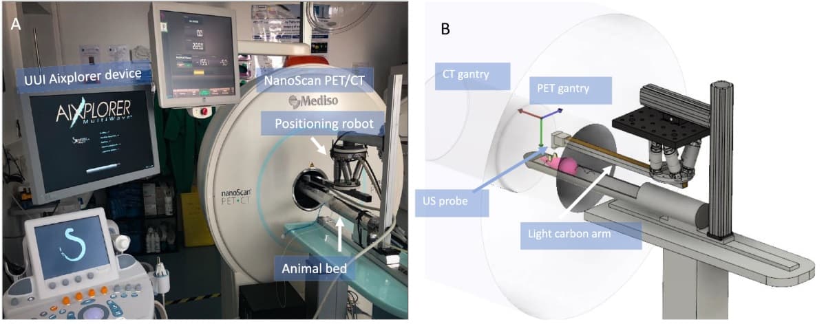
As Mailyn Pérez-Liva, a post-doctoral researcher at Inserm, explains, the PETRUS (PET registered ultrafast sonography) device combines PET, X-ray CT and ultrafast ultrasound imaging (UUI) into a single device by operating ultrasound probes in the field-of-view of a nanoScan small-animal PET/CT scanner. Photograph of the PETRUS setup (A) and a schematic representation of the PET/CT system (B). (Courtesy: M Pérez-Liva et al Phys. Med. Biol. 63 19NT01)
 Photograph of the PETRUS setup (A) and a schematic representation of the PET/CT system (B). (Courtesy: M Pérez-Liva et al Phys. Med. Biol. 63 19NT01)
Although previous work has demonstrated that the device yields “unprecedented multiparametric information for preclinical oncology and cardiology studies”, Pérez-Liva highlights the well-known fact that the presence of objects attenuating the 511 keV annihilation gamma rays inside a PET gantry may degrade image quality and create artefacts in the reconstructed images.
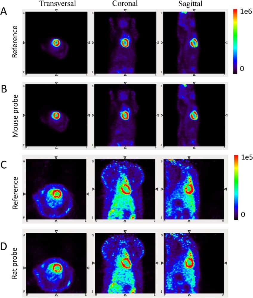
In view of the fact that the exact compositions of ultrasound probes are not provided by manufacturers, Pérez-Liva notes that the effects of their presence in the PET field-of-view cannot be reliably estimated using models. This motivated the researchers to investigate the effect on image quality experimentally and, since PET is a quantitative molecular imaging modality, to “appreciate their influence on measurements of tissue radioactivity concentrations”. FDG uptake in a mouse without (A) and with (B) the probe; and in a rat without (C) and with (D) the probe. (Courtesy: M Pérez-Liva et al Phys. Med. Biol. 63 19NT01)
 FDG uptake in a mouse without (A) and with (B) the probe; and in a rat without (C) and with (D) the probe. (Courtesy: M Pérez-Liva et al Phys. Med. Biol. 63 19NT01)
To achieve this, the team examined the effects of ultrasound probes inside the PET gantry on the quality of PET images and performed tests under the conditions described by the NEMA NU 4-2008 standard protocol for small-animal PET systems. They also investigated the effects of ultrasound probes on the quantification of in vivo dynamic studies of 18F-FDG uptake in beating mouse and rat hearts.
“We observed that the presence of a UUI probe inside the field-of-view of the nanoScan PET/CT has a minor effect on the radioactivity concentration measurements in PET images, and does not degrade significantly the quantitative and qualitative PET data derived from the images — with discrepancies below 10%,” says Pérez-Liva.
Miniaturized probes
According to Pérez-Liva, it is particularly noteworthy that custom-made ultralight probes (used to image the microvascular network in mice) had a smaller effect on image quality than commercial ones. She suggested that “careful design of next-generation miniaturized probes will render them even more stealthy”.
“With the significant advantages of simultaneous UUI and PET acquisitions, which offer the unique possibility of co-registering metabolism, vascularization, tissue elasticity and anatomy with a low-cost add-on, the PET/CT–UUI device is a remarkable means to increase the range of services offered by molecular imaging with PET,” she adds.
The PET/CT–UUI instrument was assembled from existing, commercially available devices using what Pérez-Liva describes as lightweight and portable UUI instrumentation for which dedicated, customized sequences were developed.
“Remarkably, PET/CT–UUI can produce multi-parametric information that is currently unobtainable with any other non-invasive imaging method,” she says. “PET/CT devices have been integrated in the clinic for many years, and UUI is also highly translational, since imaging modes developed for small animals can be readily applied clinically using adapted ultrasound probes. Nevertheless, UUI is a modality for which clinical applications are still in early phases of development.”
In this light, Pérez-Liva stresses that the specific clinical applications for PET/CT–UUI remain naturally speculative at this stage. But she points out that they are likely to involve a large array of organs, because UUI can be applied to any organs that are accessible with conventional ultrasound imaging.
“Typically, the deeper the organ is situated, the lower the ultrasound frequency used, and the lower the frequency, the worse the spatial resolution,” she explains. “In the worst case, the upper limit of resolution is typically in the order of 500 μm, corresponding to a 3 MHz centre frequency. This is better than the inherent resolution of PET and comparable to CT and MRI spatial resolutions.”
Andrew Williams is a freelance journalist based in Cardiff, specializing in science, technology and business.
22/10/2018 from physicsworld.com
Δεν υπάρχουν σχόλια:
Δημοσίευση σχολίου