Photosynthetic sutures promote wound healing
26 Oct 2018 Héctor Capella Monsonís
Sutures are extensively used to close wounds. However, rather than promoting active healing of the wound, they act as a passive element that provokes scar formation. In addition, the disruption of blood vessels decreases the presence of oxygen in the wound, limiting the healing. Therefore, there is growing interest in the development of bioactive sutures that promote remodelling of the tissue.
In this context, Tomás Egaña from Pontifical Catholic University of Chile, together with researchers from other South American and European universities, have developed a suture that contains genetically modified microalgae (C.reinhardtii). They showed how these microorganisms in the suture are able to produce oxygen and bioactive molecules such as growth factors, elements that promote wound regeneration (Acta Biomaterialia10.1016/j.actbio.2018.09.060).
A biofunctional “green” suture
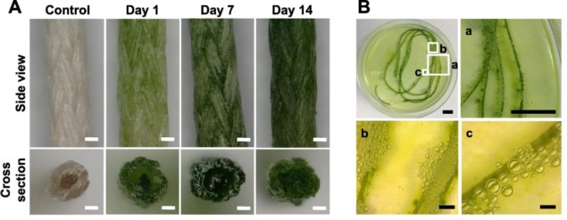
The sutures are loaded with microalgae by simply submerging them in the microorganism suspension at appropriate temperature and illumination conditions, whereby the suture’s filaments absorb the microalgae. In the study, the researchers confirmed that these organisms can proliferate within the sutures and, most importantly, carry out photosynthesis and produce oxygen. Furthermore, they also observed that human skin cells (fibroblasts) seeded on the same well as the suture can metabolize the released oxygen, showing that the seeded microalgae produced enough oxygen to be physiologically relevant. Sutures seeded with microalgae for 14 days are able to produce oxygen (bubbles) in vitro. (Courtesy: Acta Biomaterialia 10.1016/j.actbio.2018.09.060 ©2018)
 Sutures seeded with microalgae for 14 days are able to produce oxygen (bubbles) in vitro. (Courtesy: Acta Biomaterialia 10.1016/j.actbio.2018.09.060 ©2018)
To further increase the potential of this technology, the researchers investigated the possibility of the microalgae producing functional molecules that promote healing, such as growth factors. To do so, they genetically modified the microalgae to produce such factors (VEGF, PDGF and SDF-1α). For this, the investigators introduced the DNA sequences of these growth factors into the microalgae genome. They then seeded the microalgae in the sutures and observed how they produced these factors with a sustained release for 14 days. The growth factors also had biological activity on human cells in vitro.
Finally, the researchers proved that the sutures preserved their mechanical properties after hosting the microalgae for 14 days. In addition, after suturing skin for 45 times with the suture and cryopreserving (freezing at -80 °C) it, the microalgae maintained their viability and growing capacity. This demonstrates the feasibility for their repeated use and preservation.
What is next?
This technology represents a novel approach and an important advance in the development of biofunctional sutures. Also, it can be applied to other biomaterials. However, many questions remain unanswered: How feasible is the sterilization of the sutures? How do we eliminate the microorganisms once the patient is treated? How can we supply light to these photosynthetic materials? The answers to these questions and other challenges will be the focus of future efforts of Egaña and his research team.

Héctor Capella Monsonís is a PhD student contributor to Physics World. Héctor is studying biomedical engineering at the National University of Ireland, Galway. Find out more about our student contributor networks
30/10/2018 from physicsworld.com

Δεν υπάρχουν σχόλια:
Δημοσίευση σχολίου