Multimodal optical imaging ready to shine in the early detection of colorectal cancer
03 Apr 2023
Europe’s PROSCOPE R&D consortium is exploiting innovative fibre-optic technologies in tandem with endoscopic delivery to diagnose colorectal cancer in its earliest stages. Joe McEntee checks out progress towards clinical translation with project co-ordinator Peter Andersen at DTU Health Tech
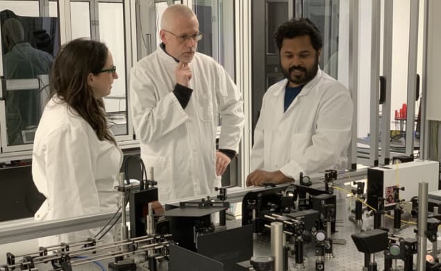
Out of the lab, into the clinic DTU Health Tech’s biophotonic imaging group is developing multimodal optical imaging techniques with the potential for at-scale clinical translation. Above: group leader Peter Andersen (centre) with colleagues Gavrielle Untracht (left) and Madhu Veettikazhy (right). (Courtesy: DTU Health Tech)
Colorectal (bowel) cancer is the second leading cause of cancer deaths in Europe – at around 160,000 deaths each year – and comes with an aggregate economic overhead in the region of €20 billion per annum – half of that relating to the primary healthcare impacts of treatment and follow-up patient care. Those figures – taken from the European Cancer Information System (ECIS), a data repository of cancer indicators and trends – reinforce the importance of early detection. When colorectal cancer is detected early (i.e. before the tumour grows beyond the colon or rectum walls into surrounding tissues), the five-year survival rate for patients increases to 90% (versus only 14% for late-stage diagnosis). Right now, though, only around four out of 10 cases of colorectal cancer are detected during that initial, localized phase of the disease.
Herein lies a clinical and commercial opportunity for biophotonics innovators to deliver early-stage, point-of-care intervention – in short, a paradigm shift when it comes to the screening, diagnosis and treatment of colorectal cancer. Think optical biopsy: the use of complementary optical imaging modalities, delivered endoscopically, to elucidate morphological, functional and molecular insights on cancerous bowel tissue without the need for excision of that tissue.
Front-and-centre within this translational R&D endeavour is Peter Andersen, group leader in biophotonic imaging at DTU Health Tech, the department of health technology at the Technical University of Denmark (DTU) in Kongens Lyngby, north of Copenhagen.
Optical opportunities
For the past three years, Andersen and his DTU Health Tech colleagues have been working to change the narrative on colorectal cancer for the better. As coordinator of the €6 million PROSCOPE initiative – funded as part of the European Union’s Horizon 2020 research and innovation programme – Andersen heads up a cross-disciplinary collaboration of scientists, industry engineers and clinicians drawn from five European countries. Their collective goal: to develop the building blocks of a multimodal fibre-optic imaging platform that paves the way for early diagnosis – at scale – of colorectal cancer, achieving specificity and sensitivity above 90% while halving the number of patients referred for excisional biopsies (which are time-consuming and prone to sampling errors that miss suspect lesions).
“Optical imaging opens up a promising pathway to earlier diagnosis and enhanced localization of disease,” explains Andersen. “By improving cancer diagnosis, we can reduce recurrence and the need for expensive follow-up procedures in the clinic – all of which means better treatment outcomes and improved quality-of-life for patients.”
Today, oncologists inspect the bowel using conventional colonoscopy systems based, for example, on high-resolution white-light video or optical narrow-band imaging. Although such approaches enable clinicians to differentiate and characterize early-stage cancerous lesions, there are limitations in terms of depth-sectioning and sensitivity.
“With this in mind,” adds Andersen, “PROSCOPE is prioritizing a unique combination of label-free [no injected dyes or biomarkers], non-ionizing and proven optical imaging modalities with a spatial resolution, specificity and sensitivity that will complement established clinical imaging procedures and reduce the need for excisional biopsy and histopathology.”
Put another way, the PROSCOPE partners are taking colonoscopy into uncharted territory. In terms of the workflow, standard white-light illumination (with on-board video camera) is used to flag up suspect lesions that merit further investigation by the clinician, at which point a portfolio of advanced optical imaging modalities comes into play. For starters, there’s optical coherence tomography (OCT), which uses low-coherence near-infrared interferometry to map reflections from different depths within tissue (i.e. depth-sectioning). Those reflections yield micron-resolution cross-sectional images of subsurface lesions in the bowel wall, also the growth of new microscopic blood capillaries that feed cancerous tissue. Peter Andersen “Optical imaging opens up a promising pathway to earlier diagnosis and enhanced localization of disease.” (Courtesy: DTU Health Tech)
 Peter Andersen “Optical imaging opens up a promising pathway to earlier diagnosis and enhanced localization of disease.” (Courtesy: DTU Health Tech)
“I liken the PROSCOPE imaging concept to the Google Earth of colonoscopies,” explains Andersen. “We start with a map of the country and then zoom into a town, then a street, then a building.” What Andersen is alluding to here is the fact that cancerous cells have a more active metabolism than adjacent, non-cancerous cells, implying higher blood flow and vessel growth to suspect lesions. As such, the PROSCOPE imaging probe uses multiphoton microscopy (MPM) to image the lesion of interest at cellular length scales – measuring blood flow, for example, and metabolic activity – while Raman spectroscopy zooms in even further to identify cancer biomarkers at the molecular level.
While the PROSCOPE partners are, for now, focusing on design iteration, miniaturization and systems integration aspects of their multimodal colonoscope, the plan is to put a technology demonstrator through its paces early next year in a small-scale clinical trial (a cohort of 20–30 patients) at the Medical University of Vienna, Austria. “The end-game for PROSCOPE is clinical translation,” says Andersen. “We’re not developing this technology because we can; we’re developing it to deliver earlier cancer diagnosis and improved patient outcomes.”
Success breeds success
Yet if PROSCOPE is all about the “what’s next” in biophotonic imaging, the consortium’s progress towards clinical application undoubtedly builds on the successes of a related, and now complete, pan-European project called MIB (Multimodal, Endoscopic Biophotonic Imaging of Bladder Cancer for Point-of-Care Diagnosis). With DTU Health Tech also the project coordinator, the MIB research consortium pioneered a cystoscope-compatible optical imaging system to enhance diagnostic capabilities with respect to bladder cancer – the ultimate goal being improved patient prognosis through early detection, earlier onset of treatment and, in turn, reduced disease recurrence (currently 50% after 12 months of follow-up).
Notable MIB outcomes include the optimization of robust and compact light sources (part of the back-end instrumentation) and the integration of multiple high-speed optical imaging probes (combining OCT, MPM and Raman spectroscopy) into a prototype cytoscopic delivery module (with biocompatible sheathing). Although follow-on R&D continues beyond the MIB framework, the consortium also took the first steps towards validation through laboratory testing (ex vivo and in vitro) and an early-stage in vivo clinical study involving 20 patients.READ MORE

“There’s a high degree of commonality across MIB and PROSCOPE,” notes Andersen. Both projects address cancers with high levels of incidence, for example, and both rely on endoscopic delivery – factors which enabled extensive knowledge transfer and a portfolio of foundational platform technologies to span the MIB and PROSCOPE programmes of work.
“More than that,” concludes Andersen, “we have also built up a granular understanding of the regulatory environment thanks to our collaboration with the Medical University of Vienna and our industry partners. In fact, PROSCOPE now forms part of a wider innovation ecosystem that will enable us to deliver advanced medical devices to meet the regulatory approvals needed for clinical translation and, ultimately, routine deployment in a diagnostic setting.”
Building the talent pipeline in biophotonics
 Optics education The 11th International Graduate Summer School in Biophotonics takes place in June on the Swedish island of Ven. (Courtesy: Peter Andersen)
Education, networking and lifelong friendships will, as in previous years, likely prove defining themes for the biomedical optics students lucky enough to make the cut for the 11th International Graduate Summer School in Biophotonics.
The school takes place in June on the small island of Ven, across the water from Helsingborg on the east coast of Sweden, and comprises a week-long programme of lectures, workshops and poster presentations. Attendees will cover a broad-scope brief, spanning the fundamental science, technology and applications of biomedical optics in diagnostic and therapeutic contexts, as well as the translation of optical modalities into life science and clinical applications.
Co-organized by DTU Health Tech’s Peter Andersen and long-time collaborator Stefan Andersson-Engels of the Tyndall National Institute at the University of Cork, Ireland, the school is restricted to a cohort of 60 PhD students and postdoctoral fellows working across diverse subdisciplines in the field of biomedical optics (with a few places allocated to talented undergraduates).
“It’s 20 years since Stefan and I organized the first summer school – a response to the noticeable void in biophotonics education and training at the time,” explains Andersen. Since then, the summer school – which is held every two years – has gone from strength to strength, attracting students and prominent guest lecturers from all the over the world.
“The school is part of the glue that knits the field of biomedical optics together,” adds Andersen. “We’re always significantly over-subscribed with early-career scientists seeking to develop their networks as well as identify future research pathways and prospective collaborators.”
For further information, see the project pages of PROSCOPE (grant agreement no. 871212) and MIB (grant agreement no. 667933).
Joe McEntee is a consultant editor based in South Gloucestershire, UK
FROM PHYSICSWORLD.COM 12/4/2023

Δεν υπάρχουν σχόλια:
Δημοσίευση σχολίου