Robotic system targets brain tumours with high-intensity ultrasound
15 Nov 2018 Tami Freeman
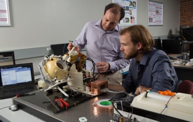
An academic–industry partnership has received a five-year, $3.5 million award from the National Institutes of Health to develop a robotic technology for minimally invasive treatment of metastatic brain tumours. The team is creating a robotic system that delivers a needle-based probe into the brain to destroy tumours with high-intensity therapeutic ultrasound. The robot is designed to operate within an MRI scanner to enable real-time treatment guidance.
The researchers, led by principal investigators Gregory Fischer from Worcester Polytechnic Institute (WPI) and Julie Pilitsis from Albany Medical College, are working closely with two corporate partners. Acoustic MedSystems will design and build the ultrasound probe and provide visualization and control software, while GE Global Research will implement thermal imaging to monitor tumour ablation in real time and help integrate the robot with a clinical MRI scanner.
“Thermal ablation has shown potential as an effective treatment, but the available devices for using this therapy have severe limitations and can’t treat all shapes, sizes and locations of tumours,” explains Pilitsis, a professor of neurosurgery. “Our hope is that this integrated robotic system will one day be able to provide all brain tumour patients with a safer, more accurate treatment.”
The robotic system, developed by the WPI research team, will align and insert a 2-mm diameter probe into the patient’s brain, via a small hole drilled in the skull, and place it within the tumour. Once in place, the probe will deliver high-intensity ultrasound energy that heats and kills tumour cells while minimizing damage to surrounding normal brain tissue. During treatment, the robot will adjust the probe’s depth and rotate it to conform the ultrasound to the shape of the tumour.
The robot will use real-time MR imaging to ensure that the probe precisely targets the tumour and to verify its position in the brain. Live MRI-based thermal imaging will monitor the dose delivered to the tumour and provide feedback on the effects of the ultrasound ablation.
“Our system is designed to provide very precise, closed-loop control,” says Fischer, professor of mechanical engineering and robotics engineering at WPI. “We will use live MR images and thermal imaging to control the pattern of the ablation and monitor and adjust it in real-time to confine the thermal effects to the area within the tumour boundaries and to ensure that we maximize the odds that we are removing the entire tumour, while minimizing the chances of damaging non-malignant tissue.”
To enable use within an MRI scanner, the robot will be made mainly from plastics and ceramics, and will use piezoelectric motors and custom motion-control electronics that generate very low levels of electrical noise, to avoid interference with the imaging system. In addition, any parts that come in contact with the patient must be sterilizable for safe operation in a surgical environment.
The WPI team is also developing a modular controller for the robot and working to integrate the robotic system with the MRI scanner, the probe’s control software and 3D navigation software. The goal is to deliver a system that can be easily integrated into the workflow inside a surgical suite.
The robotic system is an evolution of one designed and tested by the team with a previous five-year, $3 million award. “In the first phase of the project, we developed a proof-of-concept system and demonstrated that it worked as expected,” says Fischer. “With the new award, we can optimize and fully characterize the system, verify it with pre-clinical studies and get it ready for clinical use.”
15/11/2018 FROM PHYSICSWORLD.COM

Δεν υπάρχουν σχόλια:
Δημοσίευση σχολίου