Medical physics and biotechnology: our favourite research in 2022
28 Dec 2022 Tami Freeman
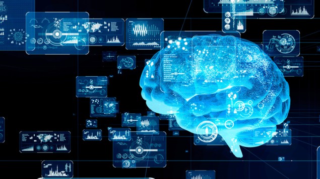
Advanced computation: Artificial intelligence techniques such as deep learning and machine learning could enhance many areas of medicine. (Courtesy: iStock/metamorworks)
From developing advanced machine-learning algorithms to building devices that will improve access to effective treatments for patients across the world, researchers working in medical physics, biotechnology and the many related fields continue to apply scientific techniques to improve healthcare worldwide. Physics World has reported on many such innovations in 2022, here are just a few of the research highlights that caught our eye.
AI in all areas
Artificial intelligence (AI) plays an increasingly prevalent role in the medical physics arena – from dealing with the vast amount of data generated during diagnostic imaging, to understanding the evolution of cancer in the body, to helping design and optimize treatments. With this in mind, Physics World hosted an AI in Medical Physics Week in June, looking at the use of deep learning for applications including online adaptive radiation therapy, PET imaging, proton dose calculation, analysis of head CT scans and identifying COVID-19 infection in lung scans.
Earlier in the year, a dedicated session at the APS March Meeting examined some of the latest medical applications of AI and machine learning, including deep learning for diagnosing and monitoring brain disorders and neurodegenerative disease, and employing AI for image registration and segmentation. Another intriguing study was EPFL’s use of a neural network to create an intelligent microscope that detects subtle precursors to rare biological events and controls its acquisition parameters in response.
The promise of proton FLASH
In a development that also made it into our top 10 Breakthroughs of the Year for 2022, this year’s ASTRO Annual Meeting saw Emily Daugherty from the University of Cincinnati Cancer Center report the findings from the first clinical trial of FLASH radiotherapy. FLASH treatments – in which therapeutic radiation is delivered at ultrahigh dose rates – hold promise for reducing normal tissue toxicity while maintaining anti-tumour activity. In this study, the researchers used FLASH proton therapy to treat 10 patients with painful bone metastases. They demonstrated the feasibility of the clinical workflow and showed that the treatment was as effective as conventional radiotherapy for pain relief, without causing unexpected side effects.
The study also represents the first-in-human use of proton FLASH. Most of the previous preclinical FLASH studies employed electrons; but electron beams only travel a few centimetres into tissue while protons penetrate far deeper. Hoping to exploit this advantage, many other groups are also investigating proton FLASH, including scientists at the University of Pennsylvania who used computational modelling to find out which is the most effective delivery technique for FLASH proton beams, and researchers from Erasmus University Medical Center, Instituto Superior Técnico and HollandPTC, who developed an algorithm that optimizes proton pencil-beam delivery patterns to maximize FLASH coverage.
Bringing back sight
Restoring vision to those who have lost the ability to see is a substantial research task. This year we reported on two studies that aim to bring this goal a step closer. Researchers at the University of Southern California are exploring the use of ultrasound stimulation to treat blindness caused by retinal degeneration. While visual prostheses that restore sight via electrical stimulation of retinal neurons have already been successfully used in patients, these are invasive devices that require complex implantation surgeries. Instead, the team demonstrated that stimulating a blind rat’s eyes with non-invasive ultrasound can activate small groups of neurons in the animal’s eye. Success story: The cornea implant restored sight to completely blind patients. (Courtesy: Thor Balkhed/Linköping University)
 Success story: The cornea implant restored sight to completely blind patients. (Courtesy: Thor Balkhed/Linköping University)
Elsewhere, a team in Sweden, Iran and India developed a new way to produce artificial corneas, using medical-grade collagen sourced from pig skin (a purified byproduct of the food industry) that the researchers chemically and photochemically treated to improve its strength and stability. In a pilot study of 20 patients, they showed that their implants were strong and resistant to degrading and could fully restore patients’ sight through minimally invasive surgery. Based on this success, Mehrdad Rafat and his team hope that the new approach could address the shortage of donor corneas for transplant and increase the treatment options for the many people worldwide in urgent need of new corneas.
Brain–computer interface innovations
Brain–computer interfaces (BCIs) provide a bridge between the human brain and external software or hardware. This year saw researchers successfully use an implanted BCI to enable a person with complete paralysis to communicate. The team – from the Wyss Center for Bio and Neuroengineering, ALS Voice and the University of Tübingen – implanted two tiny microelectrode arrays into the surface of the participant’s motor cortex. The electrodes record neural signals, which are decoded and used in an auditory feedback speller that prompts the user to select letters. The patient, who had amyotrophic lateral sclerosis (ALS) and was in a completely locked-in state with no remaining voluntary movement, learned how to alter his own brain activity according to the audio feedback received, enabling him to form words and sentences and communicate at an average rate of about one character per minute. Generating language: Neural data recorded by an implanted brain–computer interface are decoded and analysed in real time to control spelling software. (Courtesy: The Wyss Center, Geneva)
 Generating language: Neural data recorded by an implanted brain–computer interface are decoded and analysed in real time to control spelling software. (Courtesy: The Wyss Center, Geneva)
As an alternative to using implanted electrodes to sense brain activity, neural signals can also be collected non-invasively using electroencephalography (EEG) electrodes attached to the scalp. A team at the University of Technology Sydney developed a novel graphene-based biosensor that detects EEG signals with high sensitivity and reliability – even in highly saline environments. The sensor, which is made from epitaxial graphene grown on a silicon carbide-on-silicon substrate, combines the high biocompatibility and conductivity of graphene with the physical robustness and chemical inertness of silicon technology.

Tami Freeman is an online editor for Physics World
from physicsworld.com 29/12/2022

Δεν υπάρχουν σχόλια:
Δημοσίευση σχολίου