Proton CT offers low-dose treatment planning with reduced range uncertainties
16 Dec 2021 Tami Freeman
 |
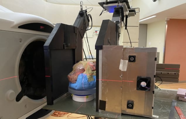
The use of protons instead of X-rays for treatment planning could improve the accuracy of proton therapy. Now a US-based research team has demonstrated that proton CT could provide low-dose planning with reduced range uncertainties, using the ProtonVDA pRAD, a prototype clinical proton imaging system.
Currently, proton therapy is planned using X-ray CT images of the patient. The CT Hounsfield units are converted into proton relative stopping power (RSP) to calculate proton range in the patient and generate the plan. This conversion, however, leads to range uncertainties and necessitates the use of margins around the tumour target. Proton CT, on the other hand, measures RSP directly, reducing uncertainties and potentially enabling smaller margins.
To investigate this potential, the team, led by researchers from ProtonVDA, Loyola University Stritch School of Medicine, Northern Illinois University and Loma Linda University, used proton CT and X-ray CT to image complex samples. They compared the directly measured proton RSP values to those determined from X-ray CT images, reporting their findings in Medical Physics.
Key comparisons
Senior author James Welsh and colleagues used a proton beamline at the Northwestern Medicine Chicago Proton Center to assess the ProtonVDA pRAD system. To validate the accuracy of proton CT-based RSP measurements, they first imaged a cylindrical phantom containing eight different tissue-equivalent inserts. Comparing the measured RSP with the known values revealed an accuracy of 1% or better for all materials bar the sinus insert (which has extremely low RSP and an absolute discrepancy of only –0.008).
Next, the researchers imaged a pig’s head and a sample of porcine pectoral girdle and ribs using the ProtonVDA pRAD and a clinical vertical X-ray CT scanner. They also scanned the head using a horizontal CT scanner at high- and low-dose settings. They then determined the differences between directly measured RSP and values from X-ray CT scans using HU to RSP conversion.
In the girdle and ribs sample, the researchers found RSP differences of 0.6% or less for all soft tissues, including muscle and adipose. However, they saw a 1.9% difference in the rib trabecular bone and a much larger difference of 6.9% in compact bone. For the pig’s head, they examined 12 regions, observing the largest RSP differences (up to 41%) in the tympanic bullae, which comprise a mixture of air, soft tissue and bone. They also noted discrepancies for the skull (up to 4.3%) and brain stem (up to 4.4%). In the eight other tissues, RSP differences ranged from –2.5% to +2.1%, with a mean of –0.4%.
The results demonstrate that although the measured and calculated RSP values are similar for soft tissues, X-ray CT could prove less accurate for treatment planning in dense bone or cavitated regions.
The team also note that proton CT delivered a far lower imaging dose (0.2–0.7 mGy for the pig’s head) than X-ray CT (3.9 and 39 mGy, for low- and high-dose scans). While the small radiation doses associated with a single scan are unlikely to ever be of harm, Welsh points out that the 10- to 100-fold reduction in dose conferred by proton CT means that such scans could be repeated regularly.
“Thus, if there is a clinical benefit to proton radiography and proton CT, the studies can be repeated as often as necessary to maximize such benefit – without the fear that such benefits will be negated by any detrimental effects from radiation dose,” he explains.
Consistency check
A proton imaging system such as the ProtonVDA pRAD can also be used to perform proton radiography prior to each treatment fraction. Comparing such images with a digitally reconstructed radiograph (DRR) from the planning CT could provide a consistency check of the X-ray CT-derived RSP map, as well as reveal discrepancies due to changes in patient anatomy or alignment.
To demonstrate this application, the researchers acquired a proton radiograph and a high-dose X-ray CT scan of the pig’s head. They then created a water equivalent thickness (WET) difference map by subtracting the calculated DRR from the proton radiograph. In most soft-tissue regions, including the brain and head-and-neck muscles, WET values for the acquired and simulated proton radiographs agreed to within 1–2 mm, equivalent to less than 1% for a total WET of up to 200 mm. The most notable differences were found in the sinus region and the tympanic bullae.READ MORE

The researchers conclude that proton CT offers potential for low-dose treatment planning with reduced margins, as well as daily pre-treatment range verification. And they have a raft of developments planned, including automating data acquisition, optical tracking of the rotation and integration with an upright treatment system. They also intend to perform more quantitative dose comparisons, and tests with treatment beams and film stacks.
“We will also be embarking on NTCP [normal tissue complication probability] studies, in which we aim to quantify the potential clinical benefits of range uncertainty reduction via proton radiography and proton CT,” Welsh tells Physics World. “In principle, reduction of range uncertainty should allow us to use tighter margins around our clinical targets and thereby reduce unwanted high dose to some normal tissues. Just how much benefit this provides will be the subject of upcoming investigations.”

Tami Freeman is an online editor for Physics World.
from physicsworld.com 1/2/2022
Δεν υπάρχουν σχόλια:
Δημοσίευση σχολίου